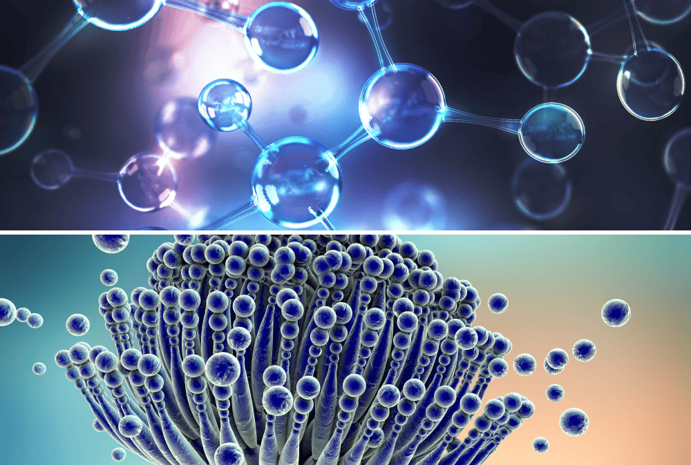
Mycotoxins are toxic metabolites produced by certain types of molds – microscopic filamentous fungi that are pervasive in both outdoor and indoor environments. Common routes of exposure to these low-molecular weight compounds include inhalation, dermal contact, and ingestion via common contaminated food sources (corn, cereals, ground and tree nuts, spices, dried fruits, apples, coffee, meat, milk, and eggs).
Attention is increasingly being given to indoor air pollution resulting not only from the influx of irritant agents (spores, pollens) from the outdoor environment, but also from the growth of molds, fungi and bacteria on almost all indoor materials (drywall, paint, wallpaper, carpeting, etc.) when excessive moisture is present in high humidity geographic areas or water-damaged buildings. The growth of these biological agents in damp environments leads to the production of spores, cells, fragments and volatile organic compounds which have been linked to a wide range of health hazards, including exacerbation of asthma as well as allergic and infectious respiratory diseases infections.
Adverse health effects may be acute or chronic in nature, and the degree of impact can vary depending on the age, sex, genetics, and underlying health status of the exposed individual, as well as the duration and dose magnitude of the offending substance and their synergistic effects with other mycotoxins.
Because mycotoxins are byproducts of mold metabolism, clinicians assessing symptomatic patients with known mold exposure – or with an environmental history concerning for mold exposure – will also need to consider the concomitant presence of mycotoxins and their potential negative health impact as well.